Human Body Care Evidence 1
Human Body Care - Evidence 1 Diego Aviña
1. Anatomical Terms
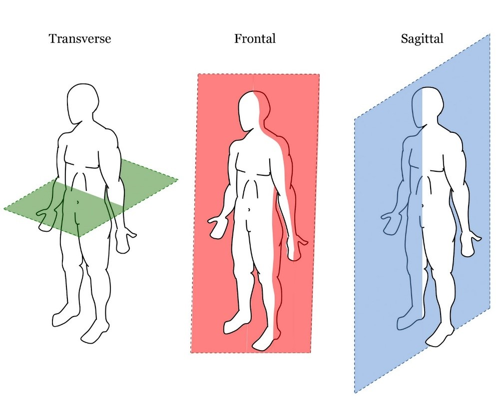
Anatomy is the science that studies body structures and the relationships among them. In the anatomical position, the person stands erect, facing the observer, with the head level and the eyes facing forward. The feet are flat on the floor and directed forward, and the arms are hanging at the sides, with the palms facing forward. We can use two terms to describe a body that is lying down: the prone position is when the body is lying face down, and the supine position is when the body is lying face up.

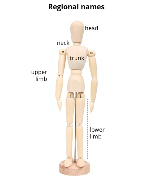
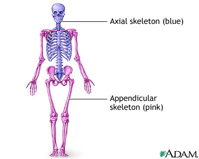
The human body can be divided into several major regions that can be identified externally, and they include the head, neck, trunk, upper limbs and lower limbs.
Directional terms are words that describe the position of one body part relative to another, and they are used to locate various body structures:
- Superior: Toward the head or upper part of a structure.
- Inferior: Away from the head or the lower part of a structure.
- Anterior: Nearer to or at the front of the body.
- Posterior: Nearer to or at the back of the body.
- Medial: Nearer to the midline (the midline is an imaginary line that divides the body into equal left and right hand sides).
- Lateral: Farther from the midline.
- Intermediate: Between two structures.
- Ipsilateral: On the same side of the body as another structure.
- Contralateral: On the opposite side of the body from another structure.
- Proximal: Nearer to the attachment of a limb to the trunk; nearer to the origination of a structure.
- Distal: Farther from the attachment of a limb to the trunk; farther from the origination of a structure.
- Superficial: Toward or on the surface of the body.
- Deep: Away from the surface of the body.
2. Description of the body tissues, components and functions of the integumentary and skeletal muscular system.
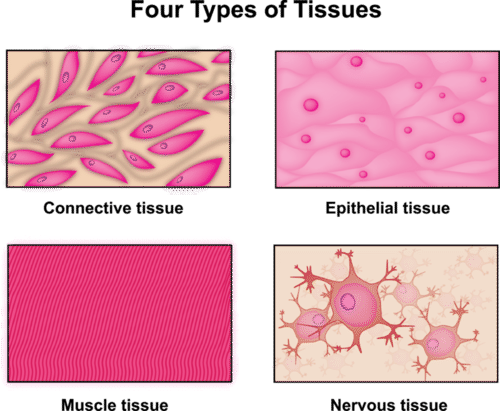
A tissue is a group of similar cells that work together to carry out a specific function. Body tissues can be classified into four basic types according to their function and structure: Epithelial tissue, connective tissue, muscular tissue and nervous tissue.
Epithelial tissue: Consists of tightly packed cells that form a continuous layer. Because the cells are closely packed, and are held tightly together by many cell junctions, there is little intercellular space between adjacent plasma membranes, epithelial tissue has many different functions in the body; the most important are protection, filtration, secretion, absorption and excretion.
Connective tissue: Is one of the most abundant and widely distributed tissues in the body. In its various forms, connective tissue has a variety of functions. It binds together, supports, and strengthens other body tissues; protects and insulates internal organs; compartmentalizes structures such as skeletal muscles; serves as the major transport system with in body (blood); is the primary location of stored energy reserves (fat); and is the main source of immune responses.
Muscle tissue: Consists of elongated cells called muscle fibers that use ATP to generate force. As a result, muscular tissue produces body movements, maintains posture, and generates heat. It also provides protection. There are three types of muscular tissue based on its location, structure and function: skeletal muscle tissue, cardiac muscle tissue and smooth muscle tissue.
Nervous tissue: Consists of two cell types, neurons and neuroglia, and is present in the brain and spinal cord. A neuron is a specialized cell that has three parts; a cell body, dendrites, and an axon.
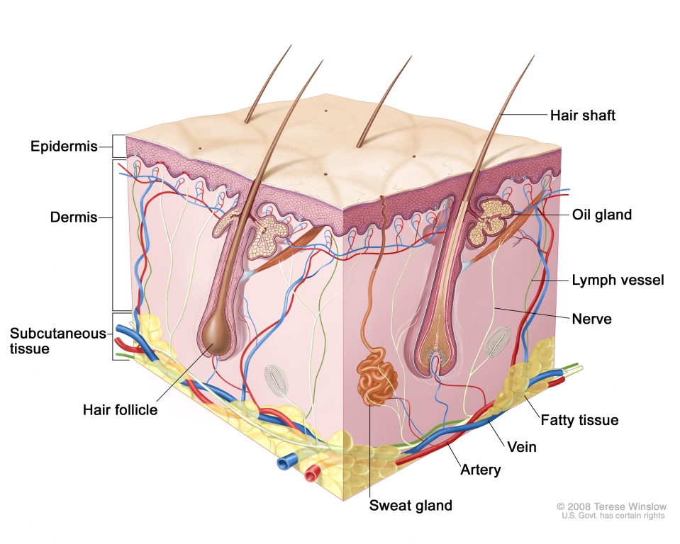
The skin has several important functions:
- Thermoregulation: the homeostatic regulation of body temperature in two ways: by liberating sweat at its surface and by adjusting the flow of blood in the dermis.
- Blood reservoir: because the dermis has an extensive network of blood vessels that carry form 8 to 10% of the total blood flow in a resting adult.
- Protection: Keratin protects underlying tissues from microbes, abrasion, heat, and chemicals and the tightly packed keratinocytes resist invasion by microbes.
- Cutaneous sensations: refers to the sensations arising from the skin, such as touch, pressure, vibration, and tickling, as well as thermal sensations such as warmth and coolness.
- Excretion and absorption: which is the elimination of substances from the body and the passage of materials from the external environment into body cells.
- Production of vitamin D: This requires the activation of a precursor molecule in the skin by UV rays in sunlight. Enzymes in the liver and kidneys then modify the activated molecule, producing calcitriol, the most active form of vitamin D.


3. Analysis of the risk factors to develop skin cancer and osteoporosis.
Skin cancer is the abnormal growth of skin cells that most often develops on skin that is exposed to the sun, although it can also occur on areas of skin that are not ordinarily exposed to sunlight. Skin cancer occurs when errors called mutations affect in the DNA of skin cells. The mutations cause the cells to grow out of control and form a mass of cancer cells. The most dangerous skin cancer cells are Melanomas, which arise from melanocytes, which produce melanin, and account for about 2% of all skin cancers. They metastasize rapidly and can kill a person within months of diagnosis.
The risk factors for developing skin cancer include:
- Fair skin
- A history of sunburns
- Excessive sun exposure
- Sunny or high-altitude climates
- Having moles or precancerous lesions know as actinic keratosis
- A personal or family history of skin cancer
- A weakened immune system
- Exposure to radiation or to certain substances such as arsenic

- Gender: Women are much more likely to develop osteoporosis than men.
- Age: The older you get, the greater your risk of osteoporosis.
- Race: Whites and those of Asian descent are at greater risk for osteoporosis.
- Family history: Having a parent or sibling with osteoporosis puts you at greater risk, especially if there is a family history of hip fracture.
- Frame size: Men and women who have small body frames tend to have a higher risk of osteoporosis because they have less bone mass to draw from as they age.
- Hormone levels: Osteoporosis is more common in people with too much or too little of the certain hormones. For example, too little estrogen or testosterone or too much thyroid or parathyroid hormone can lead to osteoporosis.
- Dietary factors: Osteoporosis is more common in people with a low calcium intake, those with an eating disorder such as anorexia, and those who have undergone gastrointestinal surgery, which tends to diminish the absorption of nutrients, including calcium.
- Lifestyle choices: People who spend a lot of time inactive have a higher rate of osteoporosis than their active counterparts. Excessive alcohol consumption interferes with the body’s ability to absorb calcium. People who use tobacco products are more likely to develop osteoporosis although the mechanism is unclear.

Hi Diego.! Your activity is complete. Well done :)
ResponderBorrar